Fraser-Kirk Plastic Surgery
Sunshine Coast
Level 3/37 The Esplanade,
Maroochydore QLD 4558
Patient profile
Age: 70s | Sex: Male | General health: Fit and well Photography: Immediately preoperative, immediately postoperative & 3 months postoperative
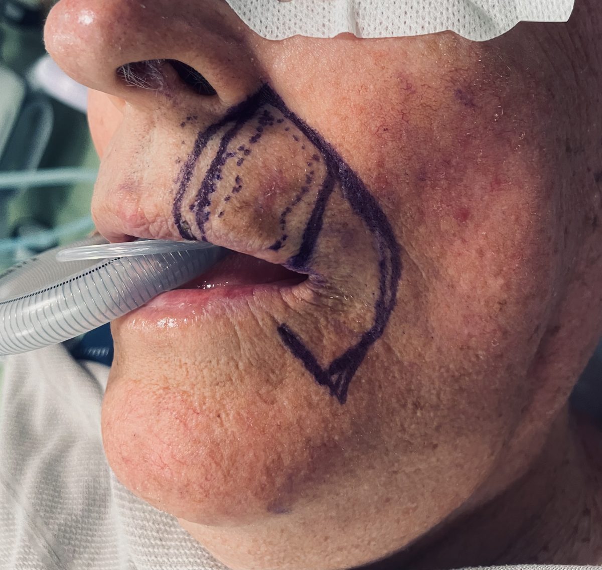
This case describes the surgical management of a melanoma in situ (lentigo maligna subtype) of the upper lip in a fit and well man in his 70s. The lesion involved the vermilion-cutaneous junction — a functionally and anatomically critical boundary — and its management required balancing the oncological priority of complete excision against the reconstructive priority of preserving a competent and mobile upper lip. The lesion presented as a variegated pigmented patch of the left upper lip extending across the vermilion-cutaneous junction. Diagnostic biopsy confirmed melanoma in situ, lentigo maligna type. The patient was otherwise fit and well, with no features to suggest invasive disease or regional spread, and the treatment goal was complete excision of the in situ melanoma with a single-stage reconstruction planned around smile symmetry and oral competence.
Detailed clinical and histopathological evaluation identified the following features:
The surgical plan was designed to achieve complete excision of the melanoma in situ while restoring a functional upper lip in a single operative episode. Two decisions defined the plan: the extent of excision, and the method of reconstruction. Excision was planned as a full-thickness wedge of the upper lip, encompassing the lesion and its vermilion-cutaneous component with a defined 7 mm peripheral surgical margin. The wedge involved approximately one third of the upper lip. This margin was chosen deliberately, and is directly supported by the evidence base for head and neck lentigo maligna. In a retrospective case series of 379 patients with head and neck lentigo maligna from Melanoma Institute Australia, a surgical margin of ≥6.5 mm (corresponding to a pathological margin of ≥3 mm) was associated with a local recurrence rate of 2.6%, compared with 27% where the margin was <6.5 mm (pathological margin <3 mm) (Crouch et al., 2021, European Journal of Surgical Oncology). My preference is for a 7 mm surgical margin, which reliably achieves a histological margin of ≥3 mm. In the anatomically constrained lip, this defined margin achieves reliable oncological clearance while avoiding an unnecessarily wide excision that would increase the reconstructive burden without demonstrated benefit. Reconstruction was planned as a Karapandzic flap — a neurovascular, musculocutaneous rotation-advancement flap that recruits the remaining orbicularis oris to reconstitute a continuous, innervated, and competent oral sphincter. To support the functional result, the plan incorporated cranial orbicularis resuspension and a caudal Z-plasty at the vermilion border.
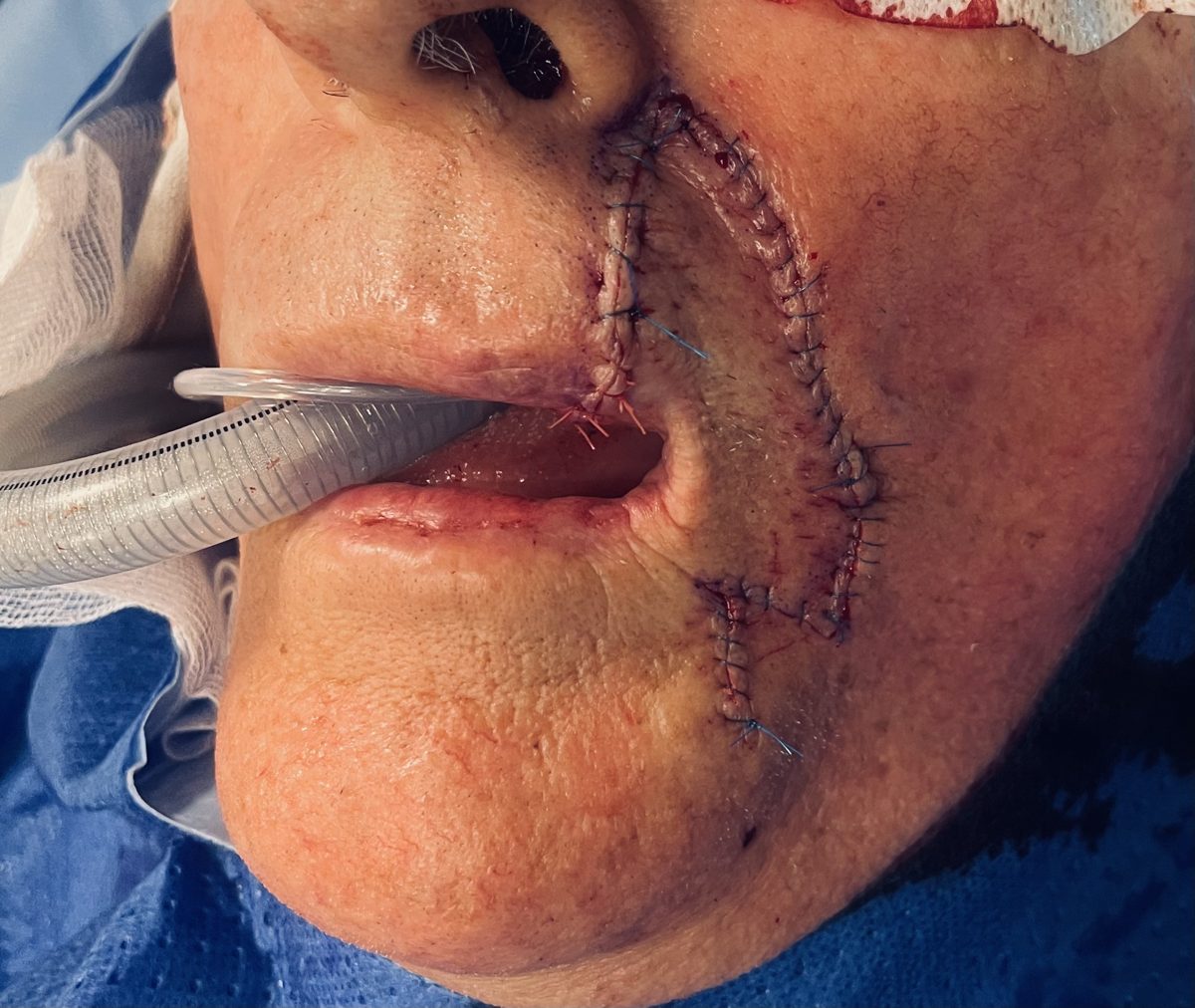
A full-thickness wedge excision of the left upper lip was performed, removing the pigmented lesion together with its vermilion-cutaneous component. The excision was taken with a 7 mm margin and encompassed approximately one third of the upper lip. Margins were oriented and marked for histopathological assessment to confirm complete excision of the in situ melanoma.
The defect was reconstructed with a Karapandzic flap. Circumoral incisions were designed to preserve the neurovascular pedicles supplying and innervating the orbicularis oris, allowing the residual muscle to be mobilised and rotated into the defect as a functional, sensate unit. This reconstitutes a continuous oral sphincter rather than simply closing the skin, which is central to maintaining oral competence and dynamic smile function.
Cranial resuspension of the orbicularis oris was performed to re-establish the vertical support of the reconstructed lip, counteracting the downward vector of the rotated flap and preserving upper lip height and philtral balance.
A Z-plasty was performed caudally at the vermilion border to break up the vertical scar line, realign the vermilion-cutaneous junction precisely, and prevent notching or a straight-line contracture across this critical boundary.
Histopathological examination confirmed that excision of the melanoma in situ was complete, with clear margins. No invasive component was identified.
At three months postoperatively, the reconstructed upper lip had healed satisfactorily, with the following features noted:
The combination of a defined, function-preserving excision margin and a neurovascular flap reconstruction achieved the dual goals of oncological completeness and functional lip preservation in a single operation.
Standardised clinical photography: immediately preoperative, immediately postoperative, and 3 months postoperative.
IMMEDIATELY PREOPERATIVE
IMMEDIATELY POSTOPERATIVE
3 MONTHS POSTOPERATIVE
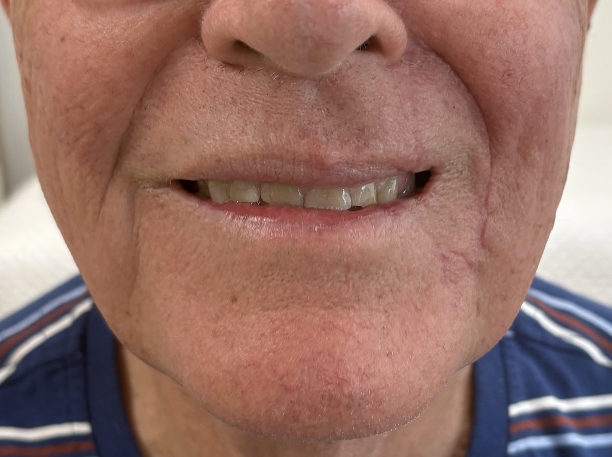 Frontal view shown. Additional views available on request.
This result is specific to this patient. It is shown with the patient’s consent and is not a prediction of any other person’s result. Outcomes vary between individuals according to anatomy, healing and other factors. These images are taken at three months, when scar maturation is still continuing and the final result has not yet settled.
| Component | Detail |
|---|---|
| Diagnosis | Melanoma in situ, lentigo maligna — left upper lip |
| Lesion site | Left upper lip, crossing the vermilion-cutaneous junction |
| Excision | Full-thickness wedge excision, 7 mm margin |
| Extent excised | Approximately one third of the upper lip |
| Margin rationale | 7 mm surgical margin (≥3 mm histological) — Crouch et al., 2021, EJSO |
| Reconstruction | Karapandzic flap (neurovascular musculocutaneous) |
| Adjunctive — cranial | Orbicularis oris resuspension |
| Adjunctive — caudal | Z-plasty at the vermilion border |
| Margin status | Complete excision; margins clear |
| Photography | Preoperative, immediately postoperative & 3 months |
The margin chosen for this excision is grounded in the largest contemporary series addressing surgical margins for head and neck lentigo maligna (melanoma in situ). In a retrospective case series of 379 patients from Melanoma Institute Australia, a surgical margin of ≥6.5 mm — corresponding to a pathological (histological) margin of ≥3 mm — was associated with a local recurrence rate of 2.6%, compared with 27% where the surgical margin was <6.5 mm (pathological margin <3 mm). On this basis, my preference for lentigo maligna of the lip is a 7 mm surgical margin, which reliably delivers a histological margin of ≥3 mm while remaining as tissue-sparing as the oncology allows in this functionally critical region. References: Crouch G, Sinha S, Lo S, Saw RPM, Lee KK, Stretch J, Shannon K, Guitera P, Scolyer RA, Thompson JF, Ch’ng S. Clinical outcomes following surgical treatment of lentigo maligna of the head and neck. Eur J Surg Oncol. 2021 May;47(5):1145–1151. doi:10.1016/j.ejso.2020.09.028. PMID: 33023795. Karapandzic M. Reconstruction of lip defects by local arterial flaps. Br J Plast Surg. 1974;27(1):93–97 — the original description of the neurovascular circumoral flap employed in this reconstruction.
AHPRA Compliance Notice: This case study is intended for general information purposes only. Individual outcomes vary, as the result experienced by one person does not necessarily reflect the result another person may experience. All surgical procedures carry risks and require a recovery period. Results shown are specific to this patient and cannot be guaranteed for others. Surgery should only be considered after thorough consultation with an appropriately qualified Specialist Plastic Surgeon, and a second opinion is encouraged. Full information on the risks of surgery and recovery is available on our risks of surgery page. This content does not constitute medical advice.
Dr David Sparks — Specialist Plastic Surgeon | FRACS (Plast.) | MED0001863770 | The Coastal Clinic, Southport QLD | Fraser-Kirk Plastic Surgery, Sunshine Coast QLD







All surgery and invasive procedures carry risks. Before proceeding, you should seek a second opinion from an appropriately qualified health practitioner. Read our full information on the risks of surgery. Dr David Sparks — Specialist Plastic Surgeon, MED0001863770.