Fraser-Kirk Plastic Surgery
Sunshine Coast
Level 3/37 The Esplanade,
Maroochydore QLD 4558
Patient profile
Age: Early 60s | Sex: Female | Relevant history: Pale complexion, history of non-melanoma skin cancers Photography: Immediately preoperative, immediately postoperative & 6 weeks postoperative
This case describes the single-stage surgical management of a basal cell carcinoma of the nasal tip in a woman in her early 60s with a pale complexion and a history of multiple non-melanoma skin cancers. The nasal tip is one of the most demanding subunits of the face to reconstruct — the skin is thick, inelastic, and often sebaceous, and small changes in contour or scar quality are visible in this central facial location. Biopsy confirmed a basal cell carcinoma with mixed infiltrative and nodular growth patterns. The infiltrative component is significant: it is a more aggressive histological subtype with a greater tendency for subclinical peripheral extension, which directly informs the excision margin. The reconstructive goal was to restore the tip contour, colour, and texture in a single operation using local tissue of matching quality, while camouflaging the resulting scar.
Detailed clinical and histopathological evaluation identified the following features:
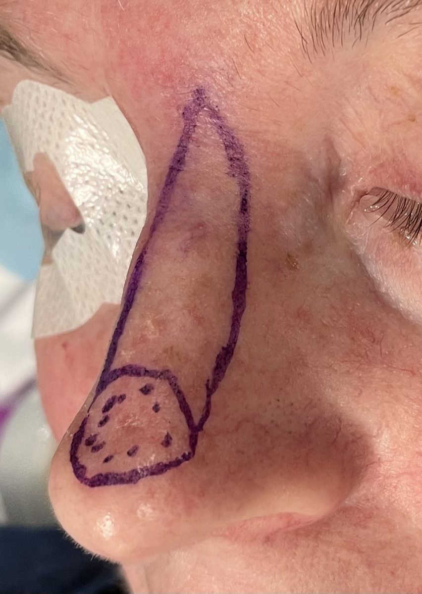
The plan was to excise the tumour with a margin appropriate to its infiltrative subtype and to reconstruct the defect in a single stage using a local island flap that recruits tissue of matched colour and contour from the nose itself. Given the mixed infiltrative and nodular histology, a 4 mm peripheral excision margin was selected, carried down to the perichondrium to ensure an adequate deep margin over the underlying cartilage framework. Reconstruction was planned as a V-Y islanded dorsal nasal (modified horn) flap based on the nasalis muscle and its axial blood supply, allowing the tip defect to be resurfaced with adjacent nasal skin in one operation and avoiding a staged forehead or interpolated flap.
Two established reconstructive principles guided the method chosen for this patient. Reconstruct ‘like with like’. Nasal tip skin differs from skin elsewhere on the face — it is thick, inelastic, and sebaceous, with a particular colour and surface texture. Replacing this tissue with tissue of the same character from the nose itself is the basis of the like-for-like principle. A skin graft, by contrast, imports thinner skin of different colour, thickness, and texture from a distant donor site, and can heal with a visible difference in contour and colour relative to the surrounding nasal skin. Recruiting adjacent nasal skin on a vascular pedicle provides a closer match of colour, contour, and texture, and does so in a single stage. Choose the incision to camouflage the scar. On the nose, a straight-line closure tends to be read by the eye as a continuous line. In this case the incision edges were planned in a deckled fashion — finely irregular, serpentine incision edges that interrupt the line of the scar so the eye does not follow it. Deckled closure is described in the reconstructive literature as a scar-camouflage method on the nose and is also used to reduce the risk of pin-cushion deformity.
This reconstruction is performed frequently in my practice for nasal tip defects, which are common on the Gold Coast, a region with among the highest skin cancer rates in the world. Recognised risks of the procedure include flap swelling (lymphoedema), which in my experience is usually transient and settles over time with massage. As with all surgery, individual results vary, and the outcome for any one patient cannot be guaranteed.
The basal cell carcinoma was excised with a 4 mm peripheral margin, carried down to the perichondrium to secure a clear deep margin over the cartilage. Margins were oriented and marked for histopathological assessment to confirm complete excision, particularly of the infiltrative component with its potential for subclinical spread.
The reconstruction employed a V-Y configuration dorsal nasal flap, designed as a horn-shaped island whose inferior border corresponds to the superior border of the defect and which extends superiorly towards the glabella. The flap recruits skin of matched colour and contour from the dorsum of the nose to resurface the tip.
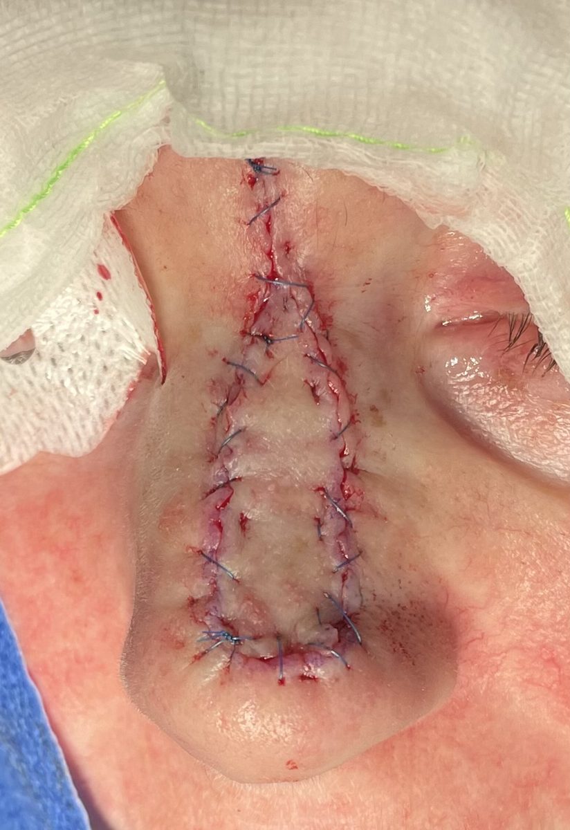
The flap was completely islanded on the nasalis muscle, preserving its axial blood supply — the angular branch of the facial artery running with the caudal nasalis. Adjacent soft tissue was dissected off the muscle and its pedicle so that the flap remained perfused entirely through this musculocutaneous vascular axis, allowing it to slide down into the tip defect with minimal tension on the muscle and its vessel.
The skin incision edges were deliberately cut in a deckled fashion — finely irregular, serpentine edges rather than a straight line. This was a considered decision: a straight incision on the nose is read by the eye as a continuous line, whereas the deckled edge interrupts that line. It is also used to reduce the risk of pin-cushion deformity and the visible apices that straight-line or W-plasty closures can leave.
The flap was advanced and inset into the nasal tip defect in a single stage, restoring the tip contour without the need for a second operation, a skin graft, or an interpolated (forehead) flap. Because the reconstructive tissue is recruited from the nose itself — a like-for-like reconstruction — the colour, thickness, and surface texture correspond closely to the surrounding nasal skin.
Histopathological examination confirmed complete excision of the basal cell carcinoma, including the infiltrative component, with clear peripheral and deep margins.
At six weeks postoperatively, the reconstruction had healed satisfactorily. It is important to note that scar maturation continues well beyond this early timepoint. Features noted included:
The combination of a margin appropriate to the infiltrative subtype and a single-stage islanded local flap achieved oncological clearance together with a reconstruction matched in colour, contour, and texture to this highly visible facial subunit.
Standardised clinical photography: immediately preoperative, immediately postoperative, and 6 weeks postoperative.
IMMEDIATELY PREOPERATIVE
IMMEDIATELY POSTOPERATIVE
6 WEEKS POSTOPERATIVE
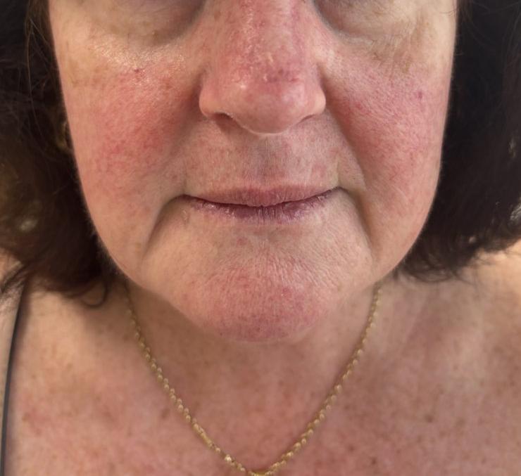 Frontal view shown. Additional views available on request.
This result is specific to this patient. It is shown with the patient’s consent and is not a prediction of any other person’s result. Outcomes vary between individuals according to anatomy, healing and other factors. These images are taken at six weeks, when scar maturation is still continuing and the final result has not yet settled.
| Component | Detail |
|---|---|
| Diagnosis | Basal cell carcinoma — infiltrative & nodular subtype |
| Lesion site | Nasal tip |
| Excision | Peripheral margin 4 mm, down to perichondrium |
| Reconstruction | V-Y islanded dorsal nasal (modified horn) flap |
| Flap pedicle | Nasalis muscle; angular branch of the facial artery |
| Flap type | Fully islanded musculocutaneous slide/advancement |
| Scar technique | Deckled incision edges for scar camouflage |
| Stages | Single-stage reconstruction |
| Margin status | Complete excision; margins clear |
| Photography | Preoperative, immediately postoperative & 6 weeks |
The reconstruction chosen for this patient reflects two established reconstructive principles — replacing nasal skin with like-for-like nasal tissue, and camouflaging the scar with deckled incision edges — each grounded in the published reconstructive literature for this region. The technique used here is based on the modified horn flap for nasal tip reconstruction: an island flap that resurfaces the nasal tip in a single-stage operation using tissue recruited from the nose itself, providing a close colour and contour match. Its vascular axis is the nasalis muscle together with its axial arterial supply, and the deckled incision edges are used specifically to camouflage the scar and reduce the risk of pin-cushion deformity. The originating series describes reliable flap perfusion following routine incorporation of the axial vessel. Reference: Deskoulidi PI, Aldred R. The modified horn flap for nasal tip reconstruction. JPRAS Open. 2021;29:26–31. doi:10.1016/j.jpra.2021.03.006.
AHPRA Compliance Notice: This case study is intended for general information purposes only. Individual outcomes vary, as the result experienced by one person does not necessarily reflect the result another person may experience. All surgical procedures carry risks and require a recovery period. Results shown are specific to this patient and cannot be guaranteed for others. Surgery should only be considered after thorough consultation with an appropriately qualified Specialist Plastic Surgeon, and a second opinion is encouraged. Full information on the risks of surgery and recovery is available on our risks of surgery page. This content does not constitute medical advice.
Dr David Sparks — Specialist Plastic Surgeon | FRACS (Plast.) | MED0001863770 | The Coastal Clinic, Southport QLD | Fraser-Kirk Plastic Surgery, Sunshine Coast QLD







All surgery and invasive procedures carry risks. Before proceeding, you should seek a second opinion from an appropriately qualified health practitioner. Read our full information on the risks of surgery. Dr David Sparks — Specialist Plastic Surgeon, MED0001863770.